Table of Contents
What is Osseous Equinus or Footballer's Ankle?
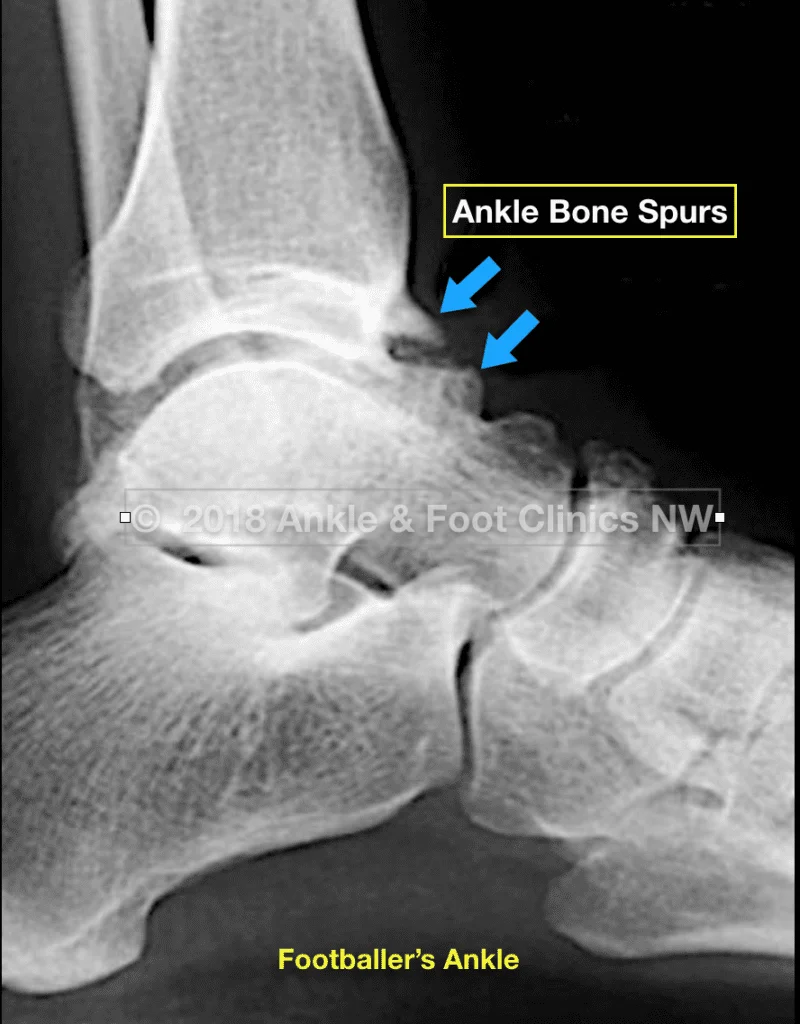
Footballers ankle is the painful build-up of bone spurs in the front of the ankle joint blocking motion. These conditions are common in athletes after repetitive ankle trauma like Soccer players. Trauma causes an increase in the joint lining, which is then pinched in the front of the ankle causing pain. The exact cause of the bone spur formation is unknown may be due to traction of soft tissues or microtrauma from repetitive kicking. Similarly a buildup of soft tissues in the ankle is called Ankle Soft Tissue Impingement .
Symptoms
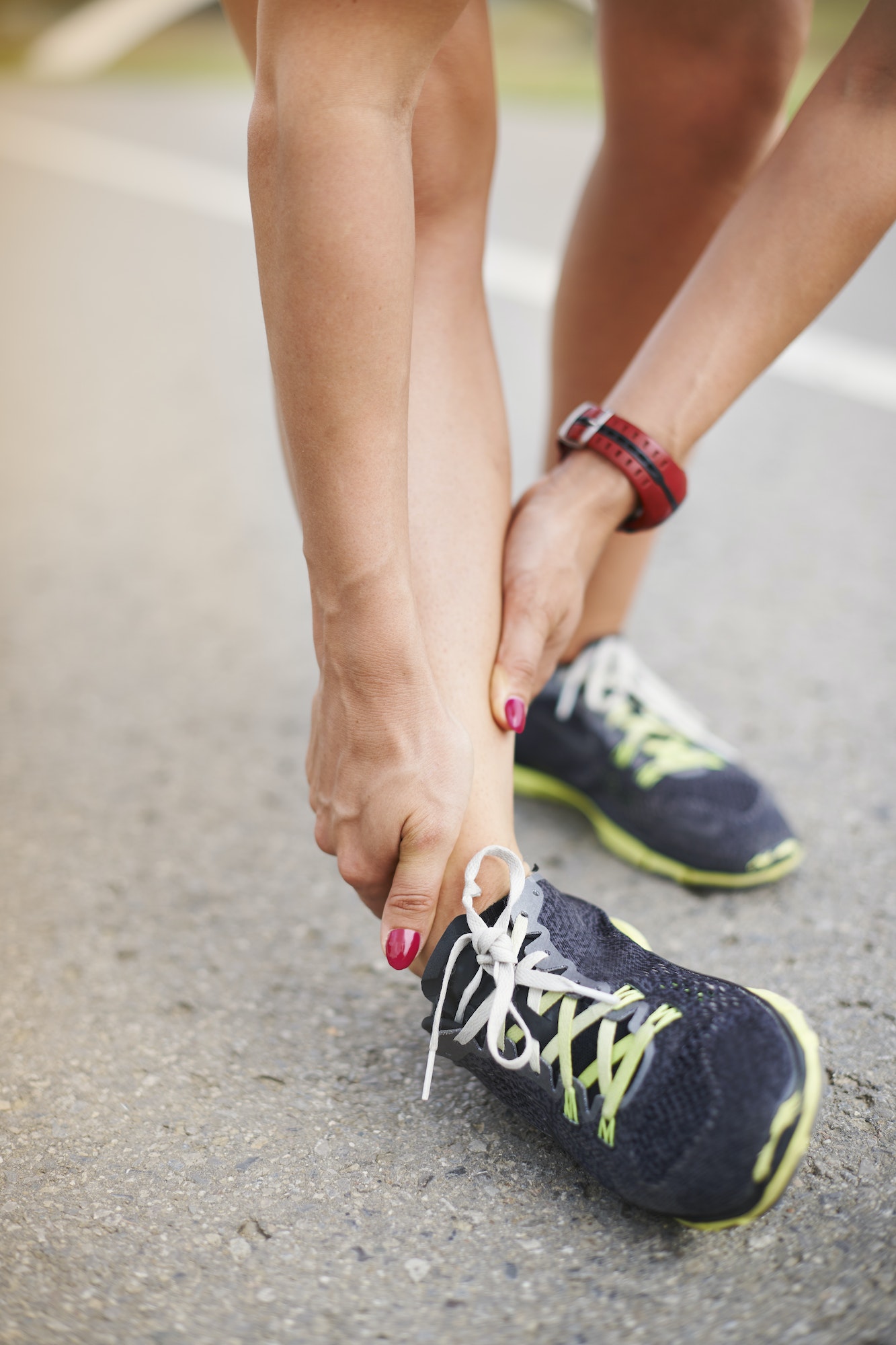
The symptoms of anterior ankle impingement are pain in the front of your ankle, usually worse with activity. There may be associated swelling and stiffness. Limited range of motion can be noticed especially when trying to bend the ankle upward towards your nose.

Diagnosis
X-rays will be taken initially. If there are bone spurs present they will show up on x-ray. Usually an MRI will also be ordered to ensure that another ankle condition such as an osteochondral lesion is not present. Ankle instability should also be ruled out and a stress exam may be recommended. Soft tissue anterior ankle impingement is largely diagnosed based upon clinical exam.
Non-Surgical Treatment
Steroid injections in the ankle can be helpful at reducing inflammation of the soft tissue. Additionally shoes with a rocker bottom can help aid in ankle range of motion during gait. Physical therapy can also be helpful.
Surgical Treatment
Arthroscopy employed by the Integrative Foot and Ankle Centers of Washington after conservative therapies have failed, can be used for the treatment of joint adhesions, bone spurring, and soft tissue build-up, which, as a result of repetitive injury, can clog the joint. It must be determined that there is no ligament instability before removing the bone spurs in the front of the ankle joint otherwise the foot can translate forward leading to further joint damage.
Typically, patients are non-weightbearing for 1-2 weeks to permit the small incisions to fully heal, slowly increasing activities as tolerated for the next 4-6 weeks, until a return to full activities can be accomplished.


